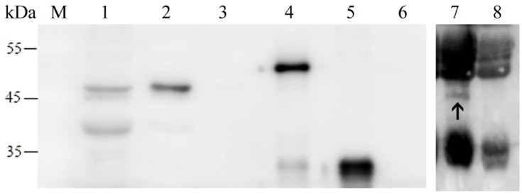
Figure 5.
Localization of MbovNase in M. bovis cells detected with western blot analysis. The different fractions of M. bovis cells, culture supernatant and the purified rMbovNase or rMbovNaseΔ181–342 were subjected to SDS-PAGE and western blot analysis with mouse antiserum to rMbovNase. MbovNase is not only present in total M. bovis cell protein (Lane 1) and membrane fraction (Lane 2), but also in culture supernatant as a secretory protein (shown by the arrow in lane 7). However, it was not in the cytoplasmic fraction (Lane 3). The rMbovNase (Lane 4) is 4 kDa larger than the nature MbovNase due to 6 ×His tag, while rMbovNaseΔ181–342 (Lane 5) is about 30 kDa, 18 kDa smaller due to the deletion of TNASE_3 domain. The pure bovine serum albumin (Lane 6) does not produce any band, while the media with many nonspecific proteins from horse serum and yeast extract display nonspecific bands in lane 7 and 8, however there is no target band in lane 8 as shown by the arrow in lane 7. Molecular weight of the reference proteins (kDa) is indicated on the left.

