Figure 1.
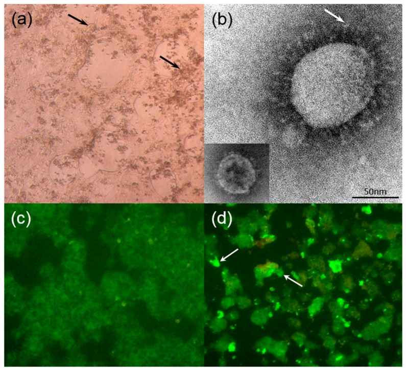
(a) HRT-18G cells infected with dromedary camel coronavirus (DcCoV) UAE-HKU23 showing cytopathic effects with rounded, aggregated, fused, and granulated giant cells rapidly detaching from the monolayer at day 5 after incubation (arrows) (original magnification 40×); (b) Negative contrast electron microscopy of ultracentrifuged deposit of HRT-18G cell culture-grown DcCoV UAE-HKU23, showing typical club-shaped surface projections (arrow) of coronavirus particles, with rabbit coronavirus HKU14 as the control (bottom left corner). Bar = 50 nm; Indirect immunofluorescent antigen detection in (c) uninfected and (d) infected HRT-18G cells using serum from dromedary showing apple green fluorescence in (arrows) DcCoV UAE-HKU23 infected HRT-18G cells (original magnification 100× for both).

