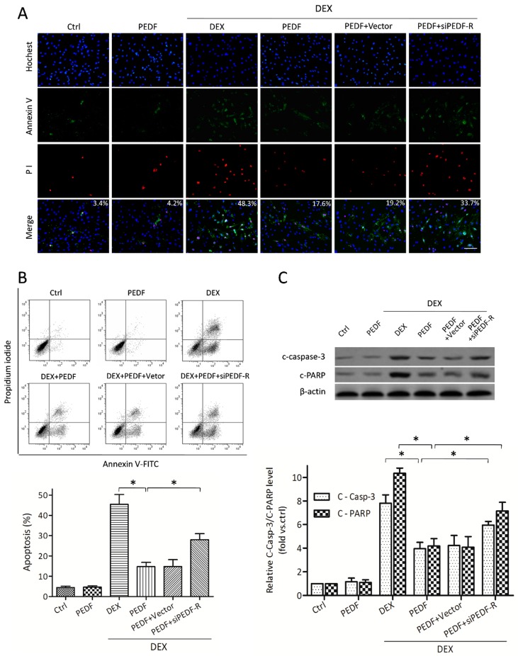
Figure 4.
The effect of PEDF on DEX-induced MC3T3-E1 pre-osteoblast apoptosis. MC3T3-E1 cells treated with or without rPEDF (10 nmol/L) were exposed to 10−5 mol/L DEX for 24 h. (A) Annexin V/PI and Hochest33342 staining assessed by fluorescence microscopy was performed to evaluate apoptosis (Annexin V+/Hochest+, mean percentage from 20 observation fields, bar = 200 μm); (B) FACS was also employed to quantify Annexin V/PI staining for apoptosis assessment. Results are expressed as apoptosis percentages (Annexin V+/Hochest+, n = 4, * p < 0.05); (C) Western blot showing the levels of cleaved caspase-3 and PARP (n = 5, * p < 0.05). Results were presented as fold induction, relative to control. Data are mean ± SD.

