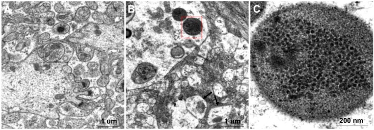
Figure 5.
Observation of the negatively stained brain tissues from RGNNV-infected and mock-infected Mandarin fish using electron microscopy. (A) Brain tissue of mock-infected mandarin fish; (B) Brain tissue of RGNNV-infected mandarin fish. Black arrows indicate that mitochondria are swollen; (C) Enlarged view from (B) shows that a lot of closely arranged particles were enclosed in the inclusion body.

