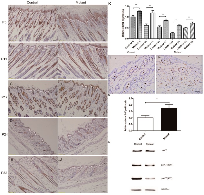
Figure 5.
Hyperproliferation of epidermis in Ppp2caflox/flox; Krt14-Cre mice and AKT signaling pathway changed. An antibody to Krt14 was used to stain tissue sections from the control (A–E) and mutant (F–J) dorsal skin at the follicular morphogenesis (P5; panels A and F), anagen (P11; panels B and G), catagen (P17; panels C and H), telogen (P24; panels D and I), and anagen (P32; panels E and J) phases. Arrows indicate a conspicuously higher number of Krt14-positive cells in the skin of mutant mice than in the epidermis of the control mice (panels A–J). All scale bars = 50 μm; (L,M) Ki67 antibody staining showed hyperproliferation in the epidermis of eight-week mutant mice epidermis compared to control littermates. All scale bars = 20 μm; (K,N) Statistical analysis indicated that the expression of Krt14 in mice skin was significantly increased at all stage. Statistical analysis indicated that the Ki67 positive cells were increased in mutant mice epidermis. n = 3 in each group, * p < 0.1, ** p < 0.01; (O) Western blot analysis of AKT, pAKT (S473), pAKT (T308), from mouse epidermis protein extracts from the knockout group and the control group. The Krt14-positive cell area was indicated by white dotted lines.

