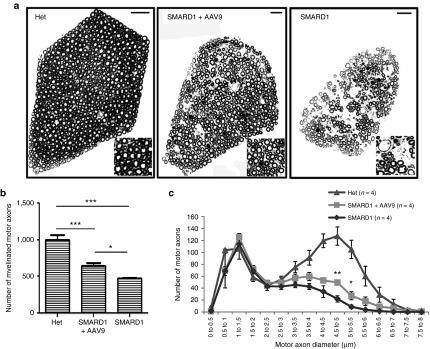
Figure 6.
AAV9-IGHMBP2 increases the total number and diameter of motor axons in 5th lumbar ventral root. (a) Entire cross sections of 5th lumbar ventral root (magnified region on the corner) of low-dose-injected males at 8 weeks of age is compared to age-matched untreated and “Het” control (n = 4 animals per group) and shows a higher density of axons in treated compared to untreated nmd that contains many areas with axonal degeneration. Scale bar = 20 m. (b) Quantification of the total number of myelinated axons demonstrates a drastic increase in the treated nmd compared to untreated (648 ± 38.54 in treated versus 471 ± 6.18 in untreated, one-way analysis of variance (ANOVA) P = 0.004; 1,000.5 ± 62.32 in “Het” versus 648 ± 38.54 in treated, one-way ANOVA P = 0.001). (c) Distribution of axonal diameter for each treatment group at 8 weeks of age. Averaged distribution of axon diameters from the entire roots of four mice for each group demonstrates that peak axonal diameter (4.5–5 µm) is significantly increased in the treated animals compared to untreated (49 ± 4.36 in treated versus 22 ± 5.27 in untreated; one-way ANOVA P = 0.007). Error bars represent mean ± SEM.

