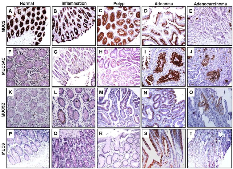
Fig. 2. Expression analysis of secretory mucins (MUC2, MUC5AC, MUC5B and MUC6) in colon disease tissue array.
Colon tissue microarrays was probed with MUC2 (EPR6145), MUC5AC (45M1), MUC5B (19.4E) and MUC6 (CLH5) monoclonal antibodies after non-specific blocking with horse serum. All sections were examined under microscope and the positive expression was evaluated on the basis of reddish brown staining by a pathologist. Representative photomicrographs are shown for MUC2 (A-E), MUC5AC (F-J), MUC5B (K-O) and MUC6 (P-T) stained tissues of normal, Inflammation, polyp, adenoma and adenocarcinoma of colon respectively. Strong expression of MUC2 observed in normal colon tissues (A). Gradual loss of expression was observed during progression to inflammation (B), hyperplastic polyps (C), adenoma (D) and adenocarcinoma (E). No immunoreactivity of MUC5AC was observed in normal colon cases (F). Expression was significantly enhanced in polyp (H), adenoma (I) and adenocarcinoma (J) of colon in comparison to normal colon (A) and inflammation cases (G). No MUC5B expression was observed in normal colon (K), while rare expression was observed during inflammation (L), polyp (M), adenoma (N) and adenocarcinoma (O). Rare expression of MUC6 was observed in cases of adenoma (S) and adenocarcinoma (T).

