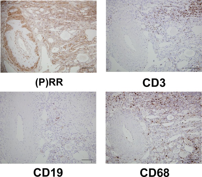
Fig 3. Staining of infiltrated cells by using (pro)renin receptor ((P)RR) and cell surface markers in serial sections.

(P)RR was positive for vessels, collecting ducts or connecting tubules, and infiltrated cells. Most of infiltrated cells positive for (P)RR were CD3-positive cells (T cell line), and CD19-positive cells (B cell line) were sparse. CD68 positive cells (monocyte/macrophage line) were diffusely scattered. Original magnification ×200. The scale bar in each figure represents 100 μm. The patient whose eGFR was 27 mL/min/1.73m2 was selected for the immunohistochemical analyses because remarkable infiltrated cells were present in the tissue section.
