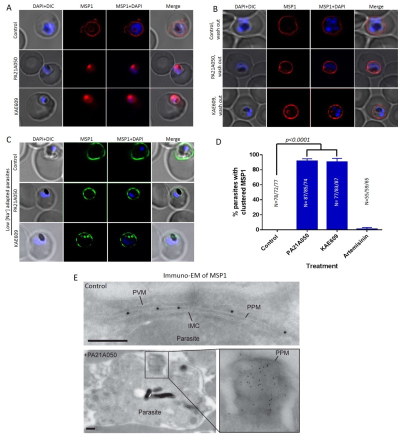
Fig 7. Reversible clustering of a GPI-anchored protein in compound treated trophozoite stage parasites.
(A) Distribution of the GPI-anchored protein MSP1 was examined by immunofluorescence assays in 32–34 h PMI (post merozoite invasion) trophozoite stage P. falciparum 3D7 exposed for 2 h to the vehicle (control), PA21A050 (10 nM) or KAE609 (10 nM). (B) After 2 h of the removal of the compounds, parasites largely restored the distribution of MSP1 throughout the PPM. (C) The compound-induced MSP1 clustering was not observed in parasites adapted to grow in low [Na+] medium following compound treatments for 2 h. (D) Quantitation of parasites showing MSP1 clustering following treatment with the indicated compound for 2 h from 3 biological replicates (the total number of parasites (N) assessed is indicated above in each treatment condition). Error bars are the SD of the percentage of clustered MSP1 parasites determined under each experimental condition (N = 3). (E) Immunogold labeling for MSP1 on a representative P. falciparum control parasite (upper panel), in which the gold particles are distributed throughout the parasite plasma membrane (PPM); and on a representative PA21A050-treated parasite (lower panel), in which the gold particles are clustered in one patch. Cryo-sections were labeled with the anti-MSP1 antibody, and binding revealed with protein A-gold particles (10 nm). PPM, Parasite plasma membrane; PVM, Parasitophorous vacuole membrane; IMC, inner membrane complex. Scale bars are 100 nm. In each immunogold labeling experiment, 60–70 parasites were imaged, and Fig 6E shows representative images.

