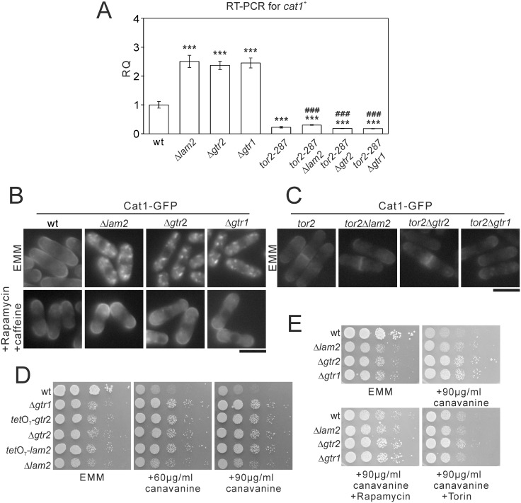
Fig 3. The deficits in Lam2, Gtr1 and Gtr2 increase the expression of cat1+ and Cat1 internalization in a TORC1-dependent manner.
(A) mRNA levels of cat1+ were increased in Δlam2 cells, Δgtr2 cells and Δgtr1 cells in a TORC1-depedent manner. The cells described in Fig 2B were grown to mid-log phase in EMM medium. Total RNA was extracted from the harvested cells and subjected to quantitative RT-PCR for cat1+ mRNA. The values were obtained by the comparative CT method in comparison to those of act1+, and then were normalized to those in wild-type cells (RQ: relative quantity). N = 3 for each group. ***P<0.001 for Turkey’s test following one-way ANOVA for the comparisons with the value of wild-type cells. ###P<0.001 for Turkey’s test following one-way ANOVA compared with respective single knockout cells. (B) Cat1 internalization in Δlam2 cells, Δgtr2 cells and Δgtr1 cells was abolished upon pharmacological inhibition of TORC1. Wild-type cells (wt, KP5859), Δlam2 cells (KP6605), Δgtr2 cells (KP6611) and Δgtr1 cells (KP6677), expressing Cat1-GFP under its native promoter were grown to mid-log phase in EMM medium. The cells were divided into two portions, one of which was treated with 0.2 μg/ml rapamycin and 10 mM caffeine for 60 min, and the other of which was left untreated. Representative fluorescent images of Cat1-GFP are shown. Scale bar, 10 μm. (C) Cat1 internalization was abolished by simultaneous tor2-287 mutation. The tor2-287 cells (KP5955), tor2-287Δlam2 cells (KP6618), tor2-287Δgtr2 cells (KP6676) and tor2-287Δgtr1 cells (KP6679) expressing Cat1-GFP under its native promoter were grown to mid-log phase in EMM medium. Representative fluorescent images of Cat1-GFP are shown. Scale bar, 10 μm. (D) The deficits in Lam2, Gtr1 and Gtr2 caused canavanine resistance. The wild-type cells (wt, KP5080), Δgtr1 cells (KP6573), tetO7-gtr2 cells (KP6645), Δgtr2 cells (KP6571), tetO7-lam2 cells (KP6650) and Δlam2 cells (KP6578) were spotted onto EMM wihout or with canavanine at 60 or 90 μg/ml. The plates were incubated at 27°C for 4 days without canavanine or for 5 days with canavanine. (E) Δlam2 cells, Δgtr1 cells and Δgtr2 cells showed canavanine resistance in a TORC1-dependent manner. The indicated cells as described in Fig 1A were spotted onto 90 μg/ml canavanine without or with rapamycin or Torin. The plates were incubated at 27°C for 4 days without canavanine or for 5 days with canavanine.

