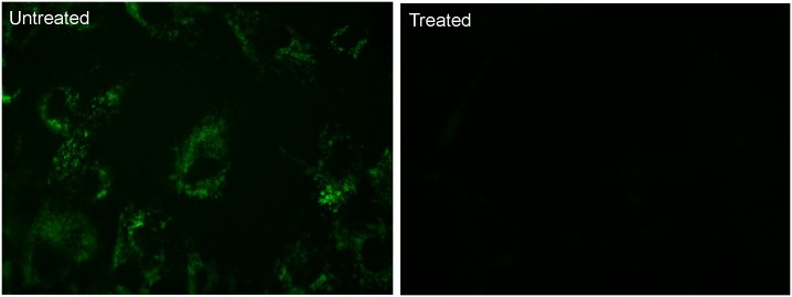
Fig 4. Inhibition of Lysosomal Metabolism by Chloroquine Leads to Reduction in Lysosomal Staining.
Healthy donor fibroblasts were incubated for 16 hours with or without 300uM chloroquine at 37°C/5% CO2. The media was removed and replaced with serum free media containing 10uM 5(6)-dimethylaminoethylcarboxamido-2',7'-dichlorofluorescein diacetate (5) and cells were incubated for 2 hours. Staining media was removed and cells were washed 3 times with PBS prior to the addition of Opti-Klear™ Live Cell Imaging Buffer. Images were then captured on a Zeiss Axio Observer A1 inverted microscope fitted with a 40X lens and FITC filter set. After treatment with chloroquine, staining in lysosomes is significantly reduced (right panel).

