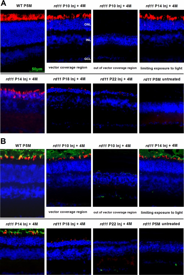
Fig 4. LPCAT1, rod rhodopsin, and cone opsin expression following AAV8 (Y733F) vector treatments on different postnatal days.
Retinal images of the posterior pole segments were collected at a distance of 0.3 mm from the optic nerve. (A) LPCAT1 immunostaining (red) of rd11 retinas at 4 months after treatment in P10 to P22-treated mice. (B) Double staining of rod rhodopsin (green) and cone opsin (red) of rd11 retinas at 4 months after treatments. Due to limited diffusion of the vector, the retinas of the P10 treated group had both rescued (vector coverage) and unrescued (out of vector coverage) areas. Age-matched wild-type C57BL/6J and untreated rd11 mice were used as controls. Nuclei were stained with DAPI (blue). P, postnatal day; Inj, injected; M, months.

