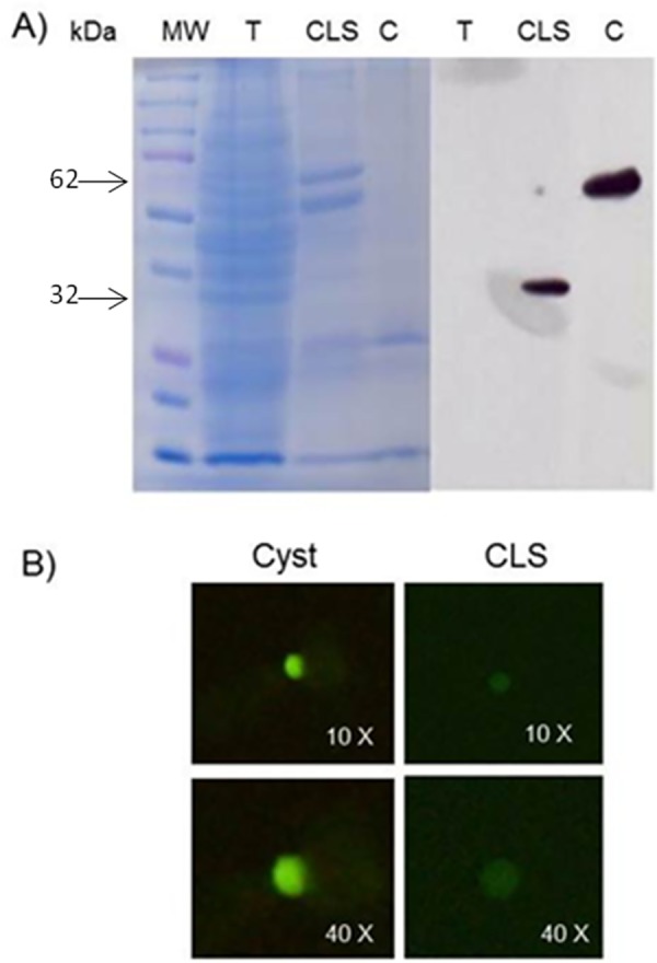
Fig 7. Jacob protein expression in CLS and cyst.

A)15 μg of sample protein were resolved on 12% acrylamide gel (Coomassie blue-stained) and transferred to a PVDF membrane. Anti-Jacob 1:1000 and a goat HRP-conjugated anti-rabbit 1:50,000 antibodies were used. Unique bands of around 30-kDa and 62-kDa were observed in CLS and cyst samples, respectively, but not in a trophozoites sample. T: Trophozoite, CLS: Cyst-like structure, C: Cyst. B) Immunofluorescence on fixed and permeabilized cyst and CLS using anti-Jacob 1:200 and a goat FITC-conjugated anti-rabbit 1:200 antibodies.
