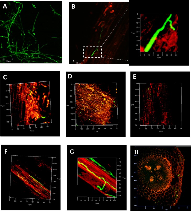
Fig 1. Early stages of chickpea root colonization by Foc 2 marked with eGFP in susceptible (JG62) cultivar by Confocal Laser Scanning Microscopy.
A. Uniform expression of eGFP in hyphae and spores of transformed isolate D4. B. Germinating conidium with primary mycelium in contact with root apex at 24 hpi. C-D. Initial hyphal colonization at lower root zone at 2 dpi. E. Intermediate root zone showing hyphal colonization extending from epidermis to cortical cells at 2 dpi. F-G. Vascular region of root getting colonized at 3 dpi. H. Fungal colonization in cortex region of DVI.

