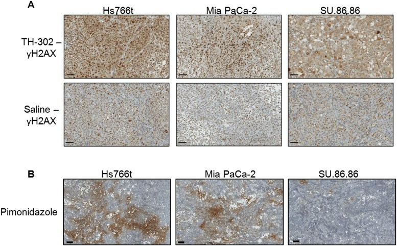
Fig 4.
(A) Histological staining of γ-H2AX, a marker for DNA damage response mechanisms, in PDAC tumors pre- and 48hr post-TH-302 treatment (50 mg/kg). Expression of γ -H2AX was increased in TH-302 treated Hs766t and Mia PaCa-2 tumors when compared to saline control. No detectable change in γ-H2AX staining was observed between Su.86.86 treated and saline tumors. (B) Pimonidazole staining as biomarker of physical tumor hypoxia. Pimonidazole Hydrochloride was injected 2hr prior to tumor removal. The extent of hypoxia was greatest in Hs766t tumors and least in SU.86.86 with Mia PaCa-2 tumors moderately hypoxic. These images are representative images of each tumor type. Scale bars = 200 μM.

