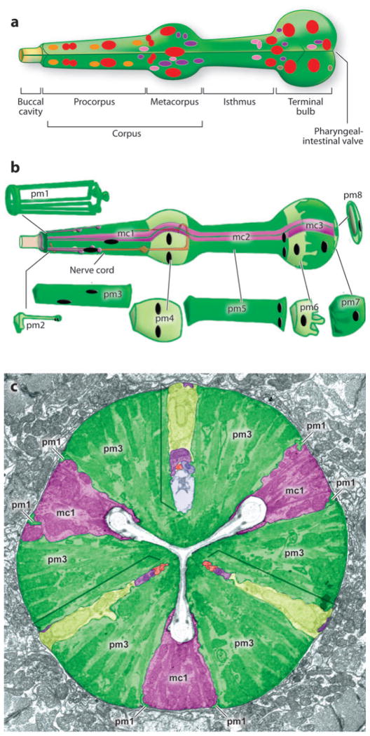
Figure 1.
Pharynx anatomy. (a) Nuclei within the pharynx are shown as muscles (red ), neurons ( purple), epithelia (orange), marginal cells ( pink), and glands (brown). Arcade cells and pharyngeal intestinal valves are not shown. (b) The bulk of the pharynx is composed of eight layers of muscles (pm1–8) ( green) and three groups of structural marginal cells (mc1–3) ( purple). (c) Muscles and marginal cells are arranged with threefold rotational symmetry, as shown in the cross section. Adapted from Mango (2007) and Altun & Hall [Wormatlas (http://www.wormatlas.org)].

