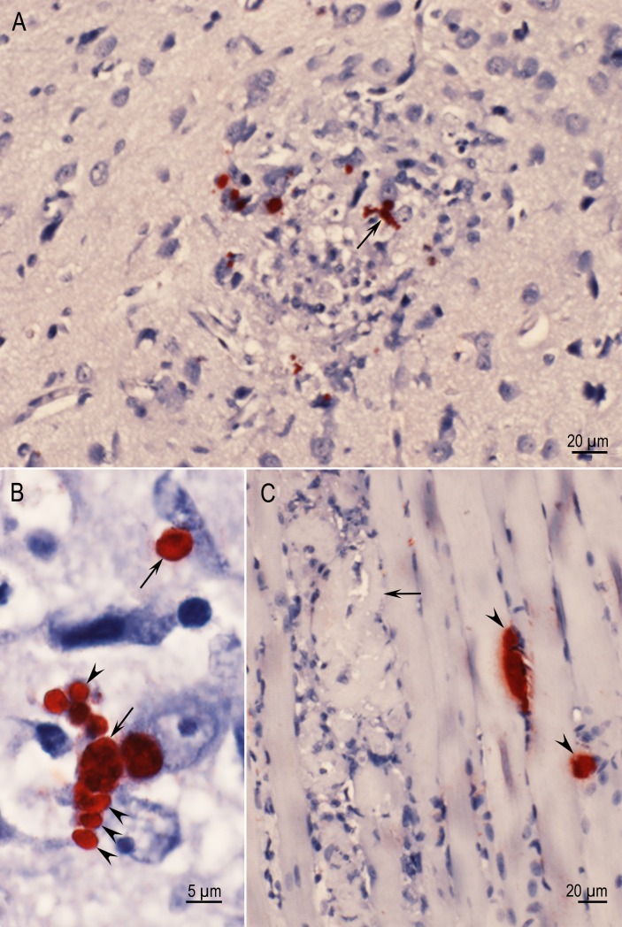
Fig 1. Acute toxoplasmosis and tissue cyst formation in cerebrum of Rat D6143 infected with the TgBbUS1 strain, 7 days p.i.
IHC staining with BAG1 T. gondii antibodies. (A) A focal area of necrosis, the genesis of a glial nodule, individual bradyzoites and small tissue cysts (arrow). Several tachyzoites are present in the lesion but are unstained. (B) Higher magnification of an area indicated by arrow in Fig 1A. Note individual bradyzoites (arrowheads) and small tissue cysts (arrows). (C) Necrosis (arrow) and two tissue cysts (arrowheads) in skeletal muscle.

