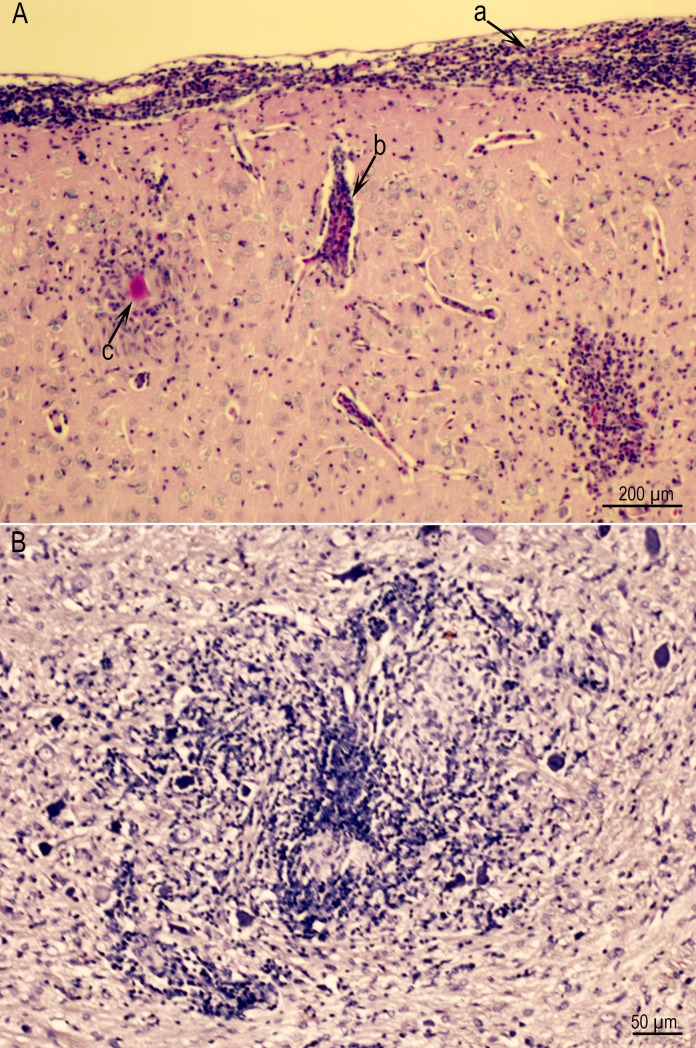
Fig 7.
(A) Meningoencephalitis in Rat D6157 infected with the TgCatCo3 strain. Note meningitis (a), perivascular infiltration of leukocytes (b), and a glial nodule with a degenerative tissue cyst (c). PASH stained. (B) A large inflammatory focus, probably resulting from the host reaction to degenerative tissue cysts in Rat D6162 infected with theGT1 strain and HE stained.

