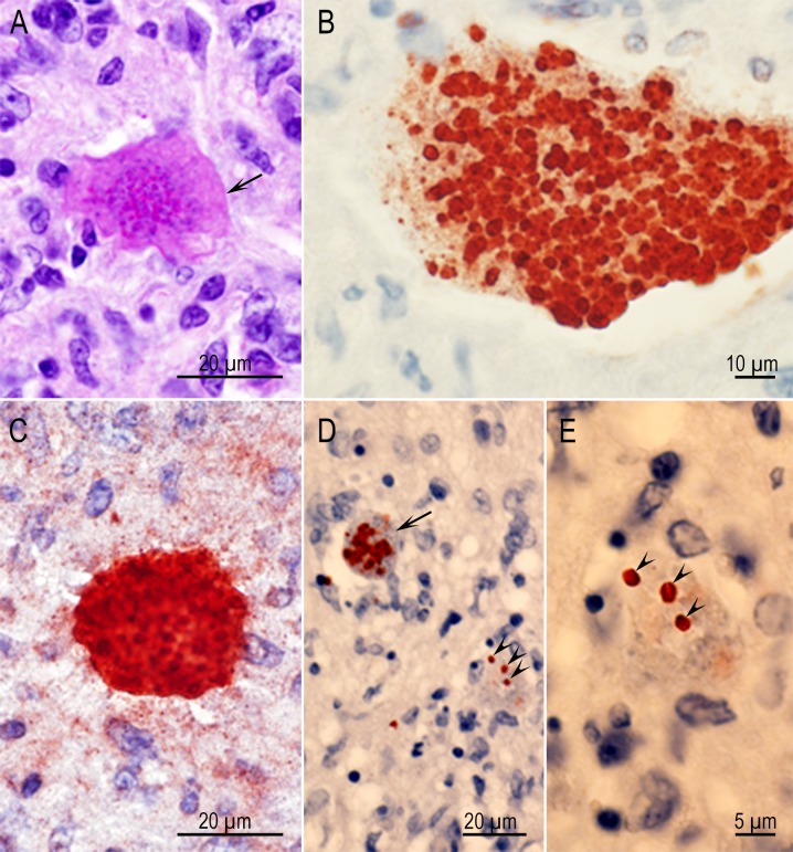
Fig 9. Examples of degenerative tissue cysts in brains of rats.
(A) Higher magnification of the lesion in Fig 7D with Rat D6157 infected with strain TgCatCo3. The cyst wall (arrow) is visible but bradyzoites have degenerated. (B) Bradyzoites in different stages of degeneration in Rat D6163 infected with the TgCatCo3 strain. The tissue cyst wall is not visible. IHC staining with BAG1 T. gondii antibodies. (C) Intensely stained bradyzoites in Rat D6162 infected with the GT1 strain. IHC staining with polyclonal T. gondii antibodies. (D, E) Two tissue cysts in different stages of degeneration in Rat D6162 infected with the GT1 strain. The outline of the tissue cysts is visible in one (arrow) and not in the other (arrowheads). The bradyzoites (arrowheads) appear to be intact.

