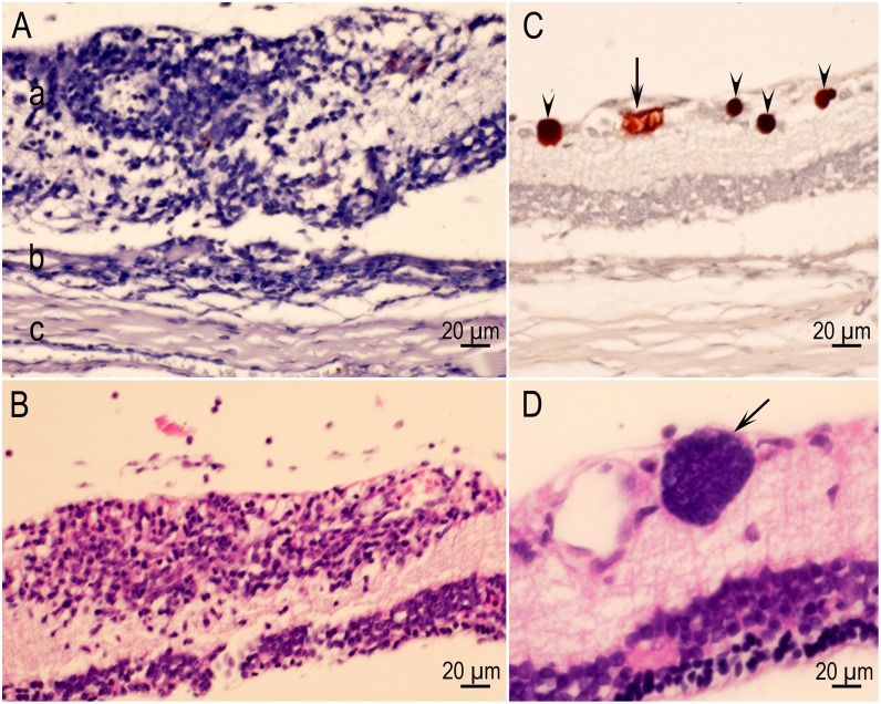
Fig 10. Retinitis in rats.
(A) Inflammatory focus involving the entire width of the retina (a) and encroaching the choroid (b). The sclera (c) is unaffected. Rat D6185 infected with the VEG strain and HE stained. (B) A focally extensive area of inflammation (retinitis) composed of lymphocytes and few macrophages affecting primarily the inner layers of the retina. Rat D6243 infected with the TgBbUS1 strain and HE stained. (C) Four intact tissue cysts (arrows) and a probable degenerating tissue cyst (arrowhead) in retina. Rat D6186 infected with the VEG strain. (D) Tissue cyst (arrow) in retina without host reaction. Rat D6249 infected with the TgBbUS1 strain and HE stained.

