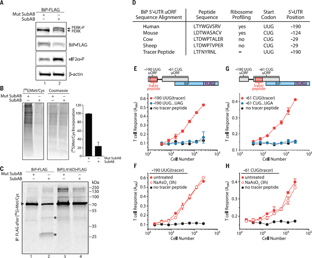
Fig. 3. The BiP 5′ UTR harbors non–AUG-initiated uORFs constitutively expressed during the ISR.
Cells treated with Mutant SubAB or SubAB (0.2 µg/ml) for 2 hours in HeLa-Kb cells were analyzed by (A) immunoblot or (B) [35S]Met-Cys pulse-labeled to measure total protein synthesis (means ± SEM; n = 4). (C) SubAB-treated HeLa-Kb cells (1 hour) followed by [35S]Met-Cys pulse-labeling (1 hour) and BiP-FLAG immunoprecipitation (►is full-length BiP and * indicates BiP cleavage products; data representative of n=2). (D) Amino acid sequence alignment of the BiP 5′ UTR −190 uORF with non-AUG start codons. (E) Schematic of the BiP 5′ UTR with the tracer peptide LYL8 at the −190 UUG uORF. Translation of the −190 UUG uORF was measured from−190 UUG uORF tracer peptide BiP-FLAG–transfected HeLa-Kb cells detected with the BCZ103 T cell hybridoma and compared with cells transfected with an identical construct containing an in-frame UAG stop codon inserted in the middle of the tracer peptide (see fig. S9) or transfected with a no-tracer peptide construct (BiP-FLAG) and (F) after treatment with NaAsO2 (10 µM) for 3 hours (means ± SD from two biological replicates and are representative of n = 3). (G) Schematic of the BiP 5′ UTR with the nested tracer peptide KOVAK in the −61 CUG uORF. Translation of the −61 CUG uORF was measured from −61 CUG uORF tracer peptide BiP-FLAG–transfected HeLa-Kb cells detected with the B3Z T cell hybridoma and compared with cells transfected with an identical construct containing an in-frame UGA stop codon inserted after the −61 CUG uORF start codon but before the tracer peptide (see fig. S9) or transfected with a no-tracer peptide construct (BiP-FLAG) and (H) after treatment with NaAsO2 (10 µM) for 3 hours (data are presented as means ± SD of two biological replicates and are representative of n = 3).

