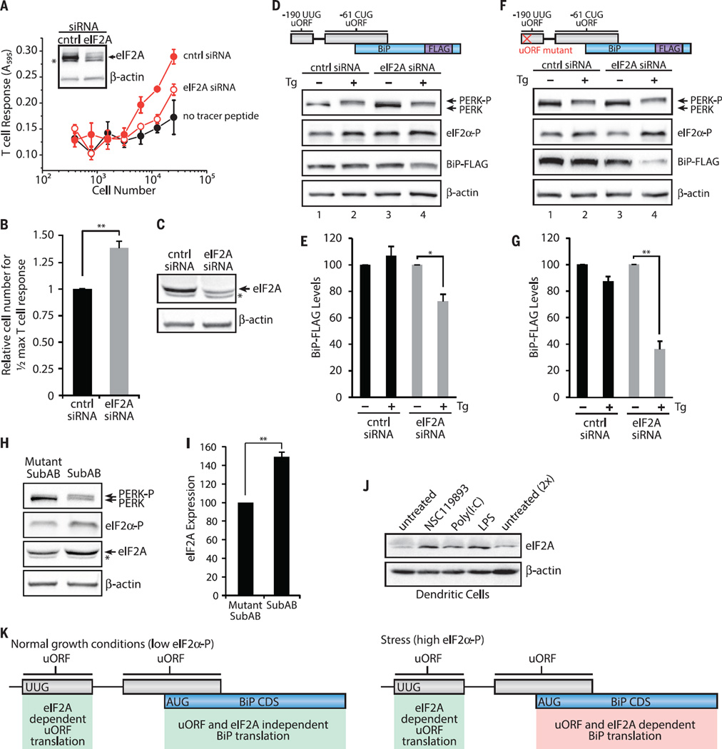
Fig. 4. eIF2A and 5′ UTR uORFs are required for sustained BiP expression during the ISR.
(A) Translation of the −190 UUG uORF from −190 UUG uORF tracer peptide (LYL8) BiP-FLAG–transfected HeLa-Kb cells after 48 hours siRNA knockdown detected with the BCZ103 T cell hybridoma and compared with cells transfected with a no-tracer peptide construct (data are presented as means ± SD of two biological replicates and are representative of n = 4). Inset shows an immunoblot for eIF2A knockdown and a β-actin loading control. (B) Relative number of cells required to achieve half-maximal T cell response for −190 UUG uORF expression with eIF2A siRNA knockdown (mean ± SEM; n = 4). Expression of BiP-FLAG analyzed by immunoblot from BiP-FLAG (D and E) or uORF mutant BiP-FLAG (F and G) transfected HeLa-Kb cells after 48 hours of siRNA knockdown (C) and treatment with DMSO or Tg (1 µM) for 3 hours (n = 3). BiP levels are presented relative to untreated for each siRNA. (H and I) HeLa-Kb cells treated with Mut SubAB or SubAB (0.2 µg/ml) for 2 hours and analyzed by immunoblot for eIF2A expression (*nonspecific anti-eIF2A antibody signal) (means ± SEM; n = 3). (J) Primary bone marrow–derived dendritic cells analyzed by immunoblot for eIF2A expression after treatment with NSC119893 (50 µM), poly(I:C) (1 µg/ml), or LPS (1 µg/ml) for 3 hours (n=2). (K) Working model for uORF translation regulation of BiP CDS expression. During normal growth conditions, BiP CDS expression is not dependent on uORF translation and eIF2A. Stress up-regulates eIF2α-P levels, leads to elevated eIF2A levels, and leads to constitutive uORF translation, which positively regulates BiP CDS expression. Statistical significance was evaluated with the unpaired t test (*P < 0.05; **P < 0.01)

