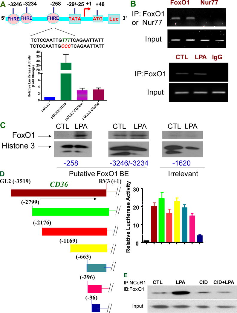
Figure 3.

FoxO1 transcriptional activities are associated with CD36 transcription. A, FoxO1 regulates activity of the CD36 promoter through recognition of FoxO1 responsive element (FHRE) at the proximal promoter. The promoter analysis shows three putative forkhead response elements (FHREs), respectively located at −258, −3234, and −3246 relative to the transcription start site. The TTT were mutated into CCC in FHRE sequence contained at proximal promoter (upper panel). Proximal promoter mutagenesis at −258 (pGL.3.2-CD36m) or deletion of proximal promoter (pGL3.2-CD36d) attenuated CD36 promoter activity (lower panel). Data represent mean ± SD of three independent experiments. B, FoxO1 binds CD36 proximal promoter (upper panel) but LPA does not enhance this binding in vivo (lower panel). HMVECs were exposed to LPA (10 μM) for 24 hrs and subjected to Chromatin immunoprecipitation analyses. Nur77 (nuclear receptor subfamily 4 group A member 1) is used as a control. Representative results are shown in triplicate experiments. C, LPA increases the interactions of FoxO1 FHRE with the CD36 proximal promoter. Based on putative binding sites for FoxO1 at CD36 promoter, biotin-labeled probes targeting FHRE respectively at proximal, distal promoter or irrelevant site were designed for detecting FoxO1 interaction with FHREs. HMVECs were exposed to LPA (5 μM) for 24 hrs, and the nuclear components were isolated and subjected to an avidin-biotin conjugated DNA binding assay. D, Distal promoter deletion does not alter CD36 promoter activity. The CD36 promoter variants in the luciferase reporter system were prepared as shown in left panel and the basal CD36 promoter activity with various deletion mutations were assayed in the HMVECs as in Figure 1A (right panel). E, PKD-1 signaling regulates recruitment of co-repressor NCoR1 to FoxO1 complex in response to LPA. HMVECs were exposed to LPA (10 μM) for 24 hrs or pre-exposed to a selective PKD inhibitor CID 755673 (25 μM) for 30 min followed with LPA (10 μM) for 24 hrs before the cell lysates were collected and subjected to co-immunoprecipitation (IP). Representative result is shown.
