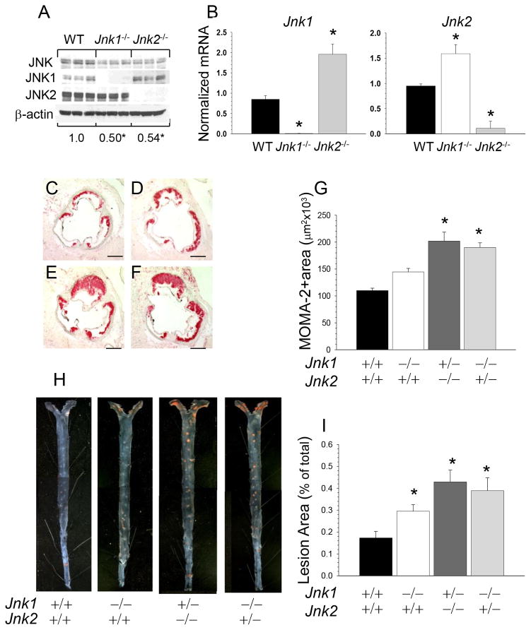
Figure 2. Genetic suppression of JNK signaling to a Jnk single allele further increases atherosclerosis.
(A) JNK protein contents in WT, Jnk1−/− and Jnk2−/− macrophages (n=3/group); Proteins were isolated and JNK protein contents were analyzed by western blot, the ratio of JNK/β-actin is presented compared to WT cells (*p<0.05 by One Way ANOVA analysis).
(B) Jnk1 or Jnk2 gene expression levels in peritoneal macrophages from mice reconstituted with WT(■), Jnk1−/−(□), or Jnk2−/−(■) FLC; mRNA levels were analyzed by real-time PCR. Graphs represent data (mean ± SEM) with the same number (n=3) of mice per group (*p<0.05 by One Way ANOVA analysis).
(C–F) Detection of macrophages in the aortic sinus lesions of mice reconstituted with WT(C), Jnk1−/−(D), Jnk1+/−/Jnk2−/−(E) or Jnk1−/−/Jnk2+/−(F) FLC. Sections were stained with MOMA-2; Scale bars, 50μm.
(G) The extent of macrophage lesion area in the proximal aorta of mice reconstituted with WT(■), Jnk1−/−(□), Jnk1+/−/Jnk2−/−(■) or Jnk1−/−/Jnk2+/−(■) FLC (*p<0.05 by One way Analysis of Variance, multiple comparisons versus control group, Tukey Test).
(H) Atherosclerotic lesions in pinned out en face aorta of mice reconstituted with WT, Jnk1−/−, Jnk1+/−/Jnk2−/− or Jnk1−/−/Jnk2+/− FLC; A pin size, 10μm.
(I) The extent of the atherosclerotic lesion area in Ldlr−/− mice reconstituted with WT, Jnk1, or Jnk1+/−/Jnk2−/− or Jnk1−/−/Jnk2+/− FLC (*p<0.05 by Kruskal-Wallis One Way Analysis of Variance on Ranks, Dunn’s Method, versus control group, WT→Ldlr−/− mice).

