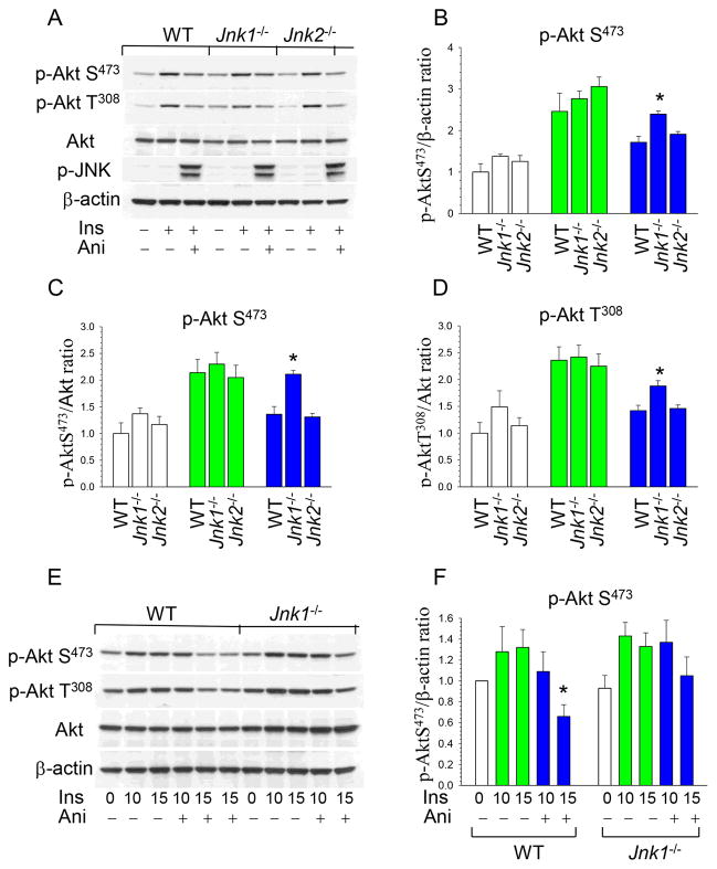
Figure 3. JNK signaling antagonizes p-Akt activity and loss of JNK1 obliterated this effect.
(A) WT, Jnk1−/− and Jnk2−/− peritoneal macrophages were pre-incubated in serum-free media for 24 hours then untreated or treated with insulin (100nM) alone or together with anisomycin (10μg/ml) for 15min. Macrophage proteins were extracted, resolved by electrophoresis (50μg), and analyzed by Western blot.
(B–D) Ratio of p-AktS473/b-actin, p-AktS473/Akt and p-Akt T308/Akt in untreated (white color) or treated with insulin (green color) or insulin plus anisomycin (blue color). Graphs represent data (mean ± SEM) of three experiments (*p<0.05 by One Way Analysis of Variance on Rank compared to control WT cells treated with insulin together with anisomycin).
(E,F) WT and Jnk1−/− macrophages were treated with insulin alone (green color) or together with anisomycin (blue color) for 10 and 15 min. Graphs represent data (mean ± SEM) of three experiments (*p<0.05 by One Way Analysis of Variance on Rank compared to WT cells treated with insulin).

