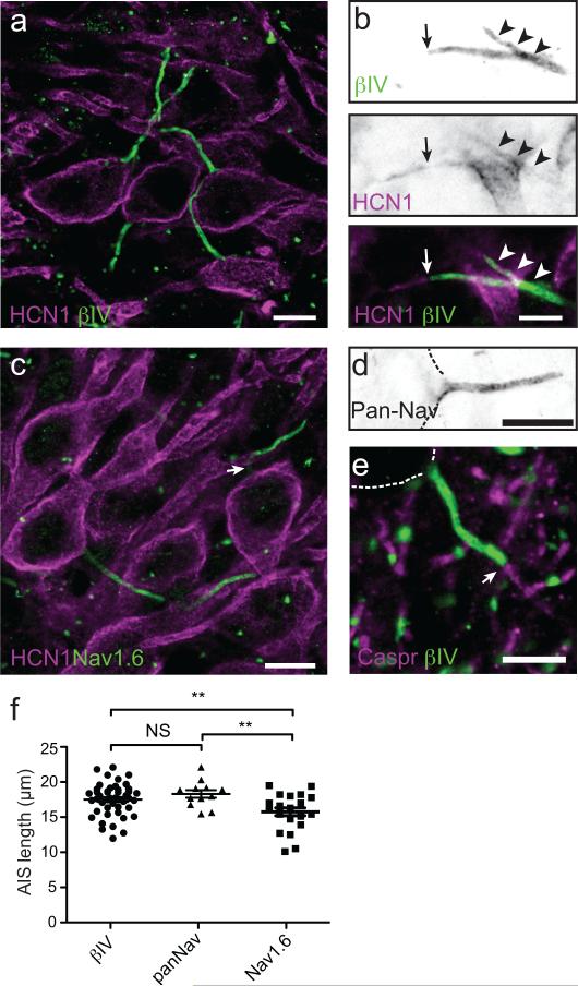
Figure 1. Expression of HCN1 subunits in the AIS of MSO principal neurons.
(a) Immunostaining of MSO neurons using antibodies against HCN1 (red) and βIV spectrin (green). Scale = 10 um. (b) Immunostaining of MSO neuron AIS using antibodies against HCN1(red) and βIV spectrin (green). Arrowheads indicate HCN1 immunoreacitivity that colocalizes with βIV spectrin. The arrow indicates the end of the βIV spectrin-labeled AIS. HCN1 immunoreactivity frequently extended beyond the end of the AIS into the more distal axon. Scale = 5 um. (c) Immunostaining of MSO neurons using antibodies against HCN1 (red) and Nav1.6. Nav1.6 immunoreactivity frequently decreased in intensity or was absent immediately adjacent to the cell body (arrow). Scale = 10 um. (d) MSO neuron AIS immunostained using a Pan-specific Nav channel antibody (PanNav). This immunoreactivity began at the transition from the cell body to the axon. Dotted lines outline the cell body. Scale = 10 um. (e) Immunostaining of MSO using antibodies against caspr (red) and βIV spectrin (green). The arrow indicates the end of the AIS and the start of the myelin sheath. Scale = 10 um. (f) length of βIV spectrin, PanNav, and Nav1.6 labeling along the AIS of MSO neurons. The length of Nav1.6-labeling was significantly less than either βIV spectrin or PanNav staining, reflecting the reduced staining near the cell body. βIV spectrin: n=46; PanNav: n=12; Nav1.6, n=22. βIV spectrin vs. PanNav: p=0.2; βIV spectrin vs. Nav1.6: p=0.006; PanNav vs. Nav1.6: p=0.001 (unpaired t-test with Welch's correction). Scatterplots show mean±SEM.

