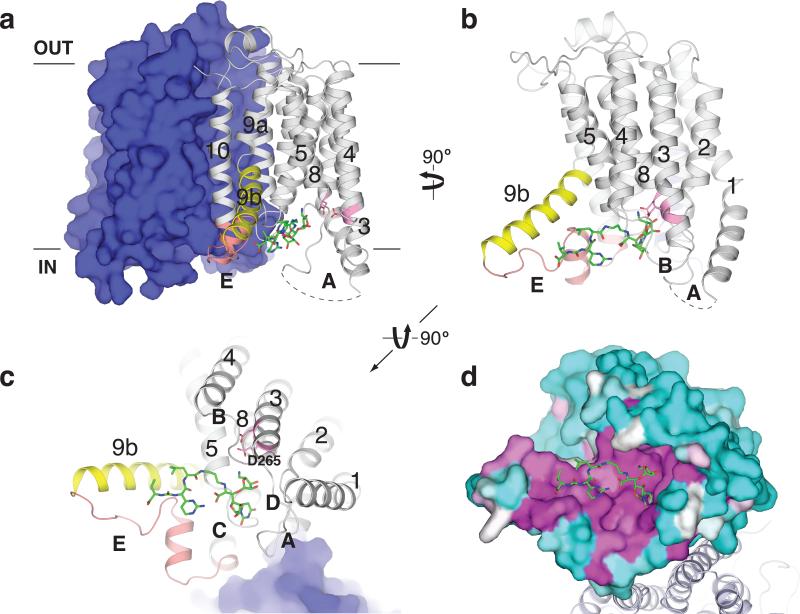
Figure 2. MD2 binds to a conserved site in MraYAA.
a, The MD2-bound MraYAA dimer viewed from the membrane. One protomer is shown as surface representation and the other as a cartoon. MD2 (green sticks) resides in the pocket formed by TMs 3-5 and 8-9b and cytoplasmic loops B-E. Conserved catalytic aspartic acid residues at the active site are shown in pink. b, View from the membrane rotated 90° about a vertical axis relative to (a). One protomer is shown for clarity. c, Cytosolic view of the MraYAA-MD2 complex, rotated 90° about a horizontal axis relative to (b). Part of MD2 is near the putative substrate recognition site formed by TM9b (yellow) and loop E (orange). d, Conservation mapping of MraYAA from high (magenta) to low (cyan) sequence identity, based on the alignment of 28 MraY homologs 21.

