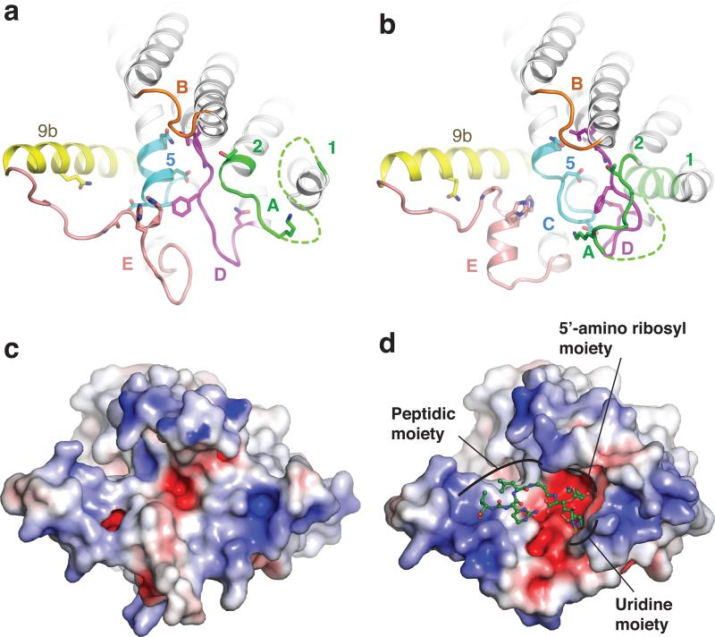
Figure 3. Conformational rearrangement of MraYAA upon MD2 binding.
a, apoMraYAA (PDB ID: 4J72) viewed from the cytoplasm, as in Fig. 2c. Residues involved in interactions with MD2 are shown as sticks. b, MD2-bound MraYAA with MD2 omitted. Part of TM1 (light green) is transparent due to its absence in the apoMraYAA structure. c, Electrostatic surface representation of apoMraYAA, viewed from the cytoplasm as in a-b. d, Electrostatic surface representation of MraYAA in complex with MD2. MD2 is green and shown in ball-and-stick representation.

