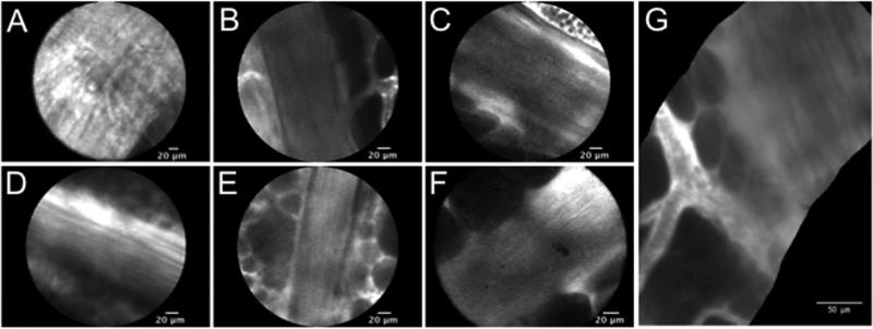
Figure 2. CLE images of the neurovascular bundle (NVB).
Nerve axons visualized with (A) the 0.85-mm probe and (B-G) the 2.6-mm probe. Nerves were visualized (B) prior to and (C-D) after NVB dissection. (E) Residual nerve structures present on the prostate capsule after neurovascular dissection. (F) Intact NVB seen ex vivo on a non-sparing prostate specimen. (G) Panoramic image of NVB generated with mosaicing algorithm from images obtained during in vivo CLE, with erythrocytes within blood vessels on the left and nerve fibers on the right.

