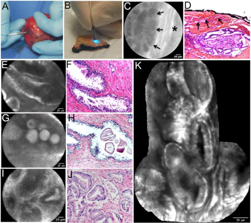
Figure 4. Ex vivo CLE imaging of prostatic tissue with corresponding hematoxylin and eosin (H&E).
(A) CLE probe application to intact prostate specimen. (B) CLE probe application to transverse cut section of prostate. (C,D) Extra capsular extension of carcinoma, arrows point to region of ECE in background of striated pattern capsule marked by * with corresponding H&E at 50× magnification (E, F) Lobular structure of benign prostatic glands. (G, H) Corpora amylacea within glands. (I, J) Prostate cancer glands with a Gleason 3+3 pattern.
Corresponding H&E for F, H and J at 100X magnification. (K) Panoramic image of normal prostate generated with a mosaicing algorithm that increased the field of view by approximately 4-fold.

