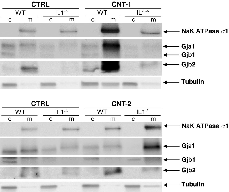
Fig. 5.
Localization of Gja1, Gjb1 and Gjb2 protein after CNT exposure. Cytosolic (c) and integral membrane/membrane-associated (m) proteins were separated into two fractions. Western blot analysis was used to investigate the localization of Gja1, Gjb1 and Gjb2. Antibodies against NaK ATPase α1 and α-Tubulin were used to detect the membrane and cytosolic fractions, respectively. The images shown are representative of two independent experiments

