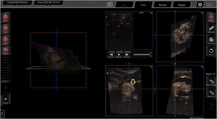
Figure 1.
Three-dimensional contrast-enhanced ultrasound done intraoperatively for completion imaging after EVAR. A type I endoleak (arrow) as seen on the Curefab CS system workstation (Curefab, Munich, Germany) that was not identified on uniplanar angiography. Reprinted from Ormesher et al. (11), Copyright 2014, with permission from Elsevier.

