Figure 7.

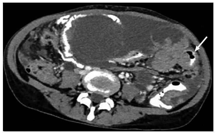
Axial computed tomography showing smooth, diffuse, eccentric bowel wall thickening involving one of the small bowel loops. Mild thickening best seen in oral contrast distended bowel loop.

Axial computed tomography showing smooth, diffuse, eccentric bowel wall thickening involving one of the small bowel loops. Mild thickening best seen in oral contrast distended bowel loop.