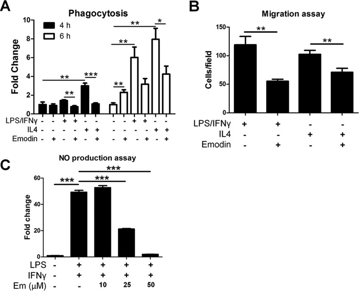
FIGURE 5.
Emodin modulates the functions of activated macrophages. Mouse peritoneal macrophages were stimulated with LPS (100 ng/ml) and IFNγ (20 ng/ml) or IL4 (10 ng/ml) with or without emodin (50 μm) for 24 h. A, macrophages were washed, and the cells were incubated with FITC-labeled E. coli bioparticles for 4–6 h. Fluorescence was detected with a microplate reader as an indicator of phagocytosis. Results are shown as the mean ± S.E. (n = 4). B, macrophages were seeded into the top chamber of a transwell insert in DMEM, and DMEM with MCP1 (20 ng/ml) was placed in the bottom of the well. After 4 h, cells were fixed, stained with DAPI, and imaged with five fields of view at ×200 magnification per membrane. Results are shown as the mean ± S.E. for two independent experiments (n = 3). C, macrophages were incubated with LPS/IFNγ with emodin (Em) at various concentrations. After 24 h, the medium was collected, and the NO content was detected. Results are shown as the mean ± S.E. (n = 4).*, p < 0.05; **, p < 0.01; ***, p < 0.001.

