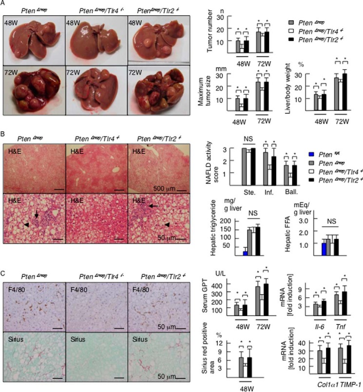
FIGURE 1.
Tlr4 deficiency delays the tumor progression and background inflammation in the PtenΔhep mice. A, representative photographs of the liver in the PtenΔhep mice, PtenΔhep/Tlr4−/− double-mutant mice, and PtenΔhep/Tlr2−/− double-mutant mice at 48 and 72 weeks of age. The tumor number, maximum tumor size, and liver to body weight ratio are shown in the right panel (48 weeks of age, n = 18–29; 72 weeks of age, n = 10 each). B, histological assessment of non-tumor areas in the liver examined on H&E staining (48 weeks of age). The PtenΔhep/Tlr4−/− mice displayed a similar grade of steatosis (upper panel), with reduced inflammatory cell infiltration (arrow) and hepatocyte ballooning (arrowhead). The non-alcoholic fatty liver disease activity score (right panel) was assessed by the grades of steatosis (Ste), inflammatory cell infiltration (Inf), and hepatocyte ballooning (Ball). Hepatic contents of triglyceride and FFA are shown in the right panel (n = 10 each). C, immunohistochemistry for F4/80 and Sirius red staining. Serum GPT levels, the gene expression of proinflammatory cytokines and profibrogenic factors in the non-tumor liver tissue, and the Sirius red-positive areas are shown in the right panel (48 weeks of age, n = 8–10). In the gene expression analysis, the fold induction is shown compared with that observed in the control Ptenfl/fl mice after normalization to the 18S RNA expression. Differences between multiple groups were compared using one-way ANOVA. The data are presented as the mean ± S.E. *, p < 0.05. NS, not significant.

