FIGURE 4.
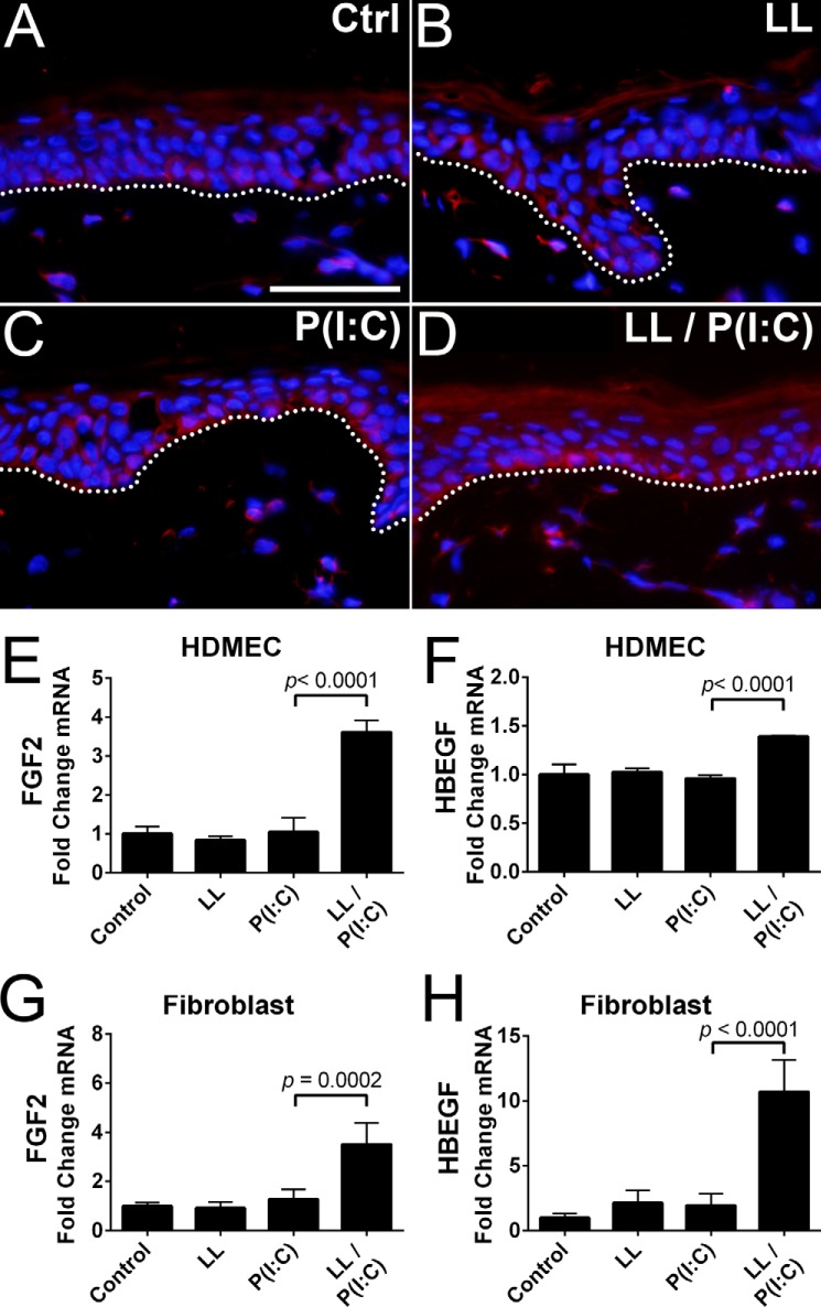
Poly(I:C) and LL-37 can stimulate FGF2 expression in basal layer keratinocytes. A–D, immunofluorescence staining of human skin punch biopsies with IgG control (IgG) antibodies indicated no background staining in basal keratinocytes. Anti-FGF2 antibodies detected low levels of FGF2 expression in basal keratinocytes of untreated control tissue (Ctrl). Tissue incubated with LL-37 (LL) or poly(I:C) (P(I:C)) had increased FGF2 expression in basal keratinocytes. Tissue co-incubated with poly(I:C) and LL-37 (LL/P(I:C)) had increased FGF2 expression compared with individual treatments. E and F, qPCR measurement of FGF2 and HBEGF from HDMECs using the treatment protocol described in Fig. 1. G and H, qPCR measurement of FGF2 and HBEGF from fibroblasts protocols described in E and F. Staining was performed on replicate punch biopsies of human skin from Fig. 2. The dotted line denotes the boundary between epidermal basal layer keratinocytes and the dermis. The scale bar represents 50 μm. p values were calculated by one-way analysis of variance. Data are mean ± S.D. of biological replicates, n = 3, and are representative data from at least three independent experiments. Error bars represent S.D.

