
FIGURE 2.
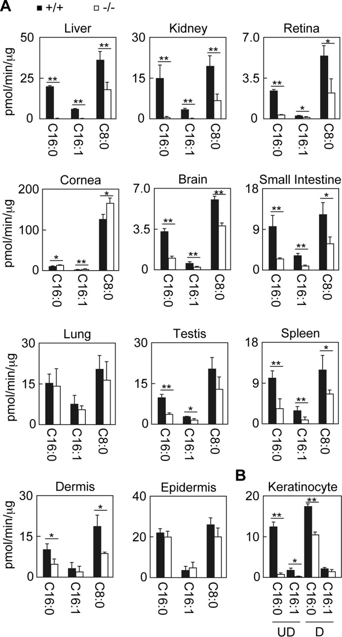
Differential contribution of Aldh3a2 to total FALDH activity depending on tissue. Total lysates were prepared from liver, kidney, retina, cornea, brain, small intestine, lung, testis, spleen, dermis, and epidermis tissue (A) and from keratinocytes kept undifferentiated or differentiated for 4 days (B) obtained from wild-type (black bars) and Aldh3a2 KO mice (white bars). Total lysates with fixed amounts of proteins (5 μg for the liver, lung, and keratinocytes; 10 μg for the kidney, cornea, small intestine, testis, spleen, epidermis, and dermis; and 15 μg for the brain and retina) were incubated with 500 μm NAD+ and 100 μm hexadecanal (C16:0), trans-2-hexadecenal (C16:1), or octanal (C8:0) for 15 min (liver, kidney, small intestine, lung, testis, and spleen) or 30 min (retina, cornea, brain, dermis, epidermis, and keratinocytes) at 37 °C. The amount of NADH product was determined by measuring the fluorescence of NADH using an Infinite M200 monochromator. Values represent the means ± S.D. of three independent experiments. Statistically significant differences are indicated (**, p < 0.01; *, p < 0.05; Student's t test). UD, undifferentiated keratinocytes; D, differentiated keratinocytes.