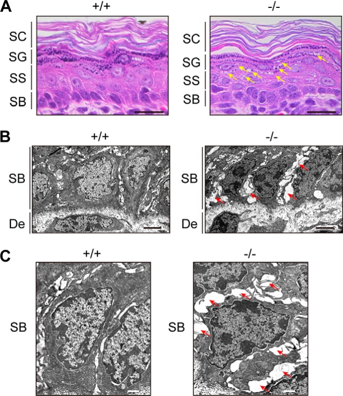
FIGURE 7.

Vacuolization and broadened intercellular spaces present in Aldh3a2−/− epidermis. A, paraffin sections (4 μm) of skin prepared from wild-type and Aldh3a2 KO mice at P0 were subjected to H&E staining. Bright field images were photographed under a DM5000B light microscope. Arrows indicate vacuolization. Bar, 20 μm. B and C, skin sections of wild-type and Aldh3a2 KO mice at P0 were subjected to electron microscopy. Arrows indicate broadened intercellular spaces. Bar, 3 μm (B), 1 μm (C). SC, stratum corneum; SG, stratum granulosum; SS, stratum spinosum; SB, stratum basale; De, dermis.
