
FIGURE 1.
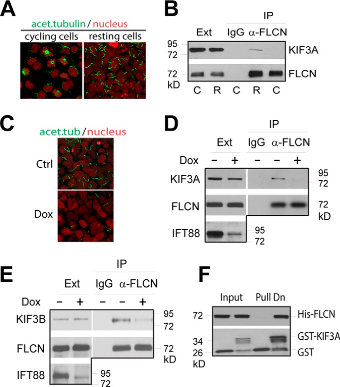
FLCN interacts with kinesin-2. A, HKC-8 cells were harvested before (cycling) or 3 days after cell density reaching confluence (resting). Cells were stained with anti-acetylated tubulin antibody (green) and nuclei with sytox (red) and examined with confocal microscopy. B, extracts from cycling (C) and resting (R) cells were immunoprecipitated with anti-FLCN antibody (α-FLCN) or control IgG. The levels of KIF3A and FLCN in the cell extracts (Ext) and precipitates (IP) were analyzed by Western blotting. C, HKC-8 cells stably expressing a doxycycline inducible IFT88 shRNA were grown in the presence (Dox) or absence (Ctrl) of doxycycline until 3 days after cell density reaching confluence. The ciliation of the cells was shown by fluorescent imaging as in A. D, extracts from cells treated as in C were immunoprecipitated with anti-FLCN antibody (α-FLCN). The levels of KIF3A, FLCN and IFT88 in the extracts (Ext) and precipitates (IP) were examined by Western blotting. E, extracts from cells treated as in C were precipitated with anti-FLCN antibody (α-FLCN) or control IgG. The levels of KIF3B and FLCN in the extracts (Ext) and precipitates (IP) were analyzed by Western blotting. F, purified GST or GST fused C-terminal globular domain (amino acids 597–701) of KIF3A (GST-KIF3A) was incubated with purified His-tagged full length FLCN (His-FLCN). The amounts of His-FLCN co-purified with GST or GST-KIF3A (Pull Dn) were analyzed by Western blotting.