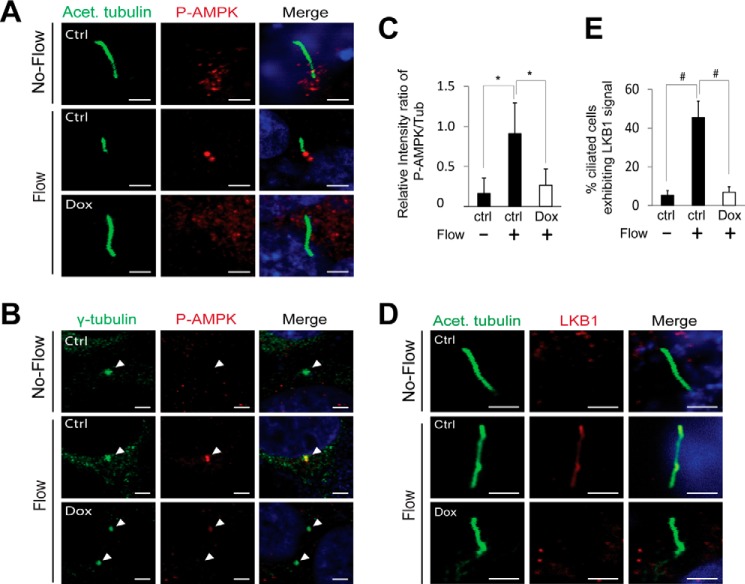
FIGURE 5.
FLCN is required for ciliary localization of LKB1 and AMPK activation at basal bodies. HKC-8 cells stably expressing doxycycline inducible FLCN shRNA were grown in the absence (Ctrl) or presence (Dox) of doxycycline under flow or no-flow condition. A, cells were stained with anti-phospho-AMPK antibody (red), anti-acetylated tubulin (green) antibody that marked primary cilia, and DAPI (blue). B, cells were stained with anti-phospho-AMPK antibody (red), anti-γ tubulin antibody (green) that marked basal bodies, and DAPI (blue). C, quantitative presentation of phosphorylated AMPK shown in B. The density of the antibody staining at the basal bodies was analyzed with Image J (NIH) software. The relative levels of phosphorylated AMPK at the basal bodies were expressed as the ratio between the fluorescent intensities of phospho-AMPK and tubulin staining. A total of 100 pairs of basal bodies from three independent experiments performed under identical experimental settings was analyzed for each indicated condition. *, p < 0.01. D, cells were stained with anti-acetylated tubulin antibody (green), anti-LKB1 antibody (red), and DAPI (blue). The stained cells were imaged by confocal microscopy. Scale bar, 5 μm. E, percentage of the ciliated cells displayed detectable LKB1 under the indicated conditions was determined. A total of 200 ciliated cells from three independent experiments performed under identical experimental settings was examined for each condition. #, p < 0.001.

