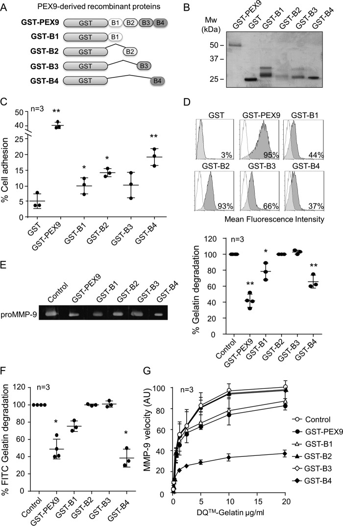
FIGURE 4.
Blades B1 and B4 of PEX9 inhibit gelatin degradation by MMP-9. A, schematic drawing of the truncated recombinant GST fusion proteins. B, SDS-PAGE analysis of the purified proteins shown in A. C, BCECF-AM-labeled MEC-1 cells were added to wells coated with the indicated proteins (0.4 μm), and adhesion was quantitated as explained. D, MEC-1 cells were incubated for 30 min with or without the indicated proteins (0.8 μm) and analyzed by flow cytometry using anti-GST antibodies. E, representative gelatin zymography analysis of 20 ng of purified recombinant proMMP-9 in the presence or absence of the indicated proteins. The values (arbitrary units) represent the averages of three different experiments. F, 60 nm of MMP-9 was added to FITC-gelatin-coated plates in the absence or presence of the indicated proteins. After 24 h, the fluorescence was determined. G, effect of the indicated proteins on the conversion of DQ-gelatin into fluorogenic gelatin. The graph represents the maximal enzyme velocity evolution as a function of the amount of substrate. *, p < 0.05; **, p < 0.01. AU, arbitrary units.

