
FIGURE 1.
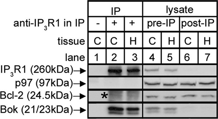
Co-IP of Bok with IP3R1 in mouse brain tissues. Mouse brain cerebral cortex (C) and hippocampus (H) lysates were incubated without or with anti-IP3R1, and IPs (lanes 1–3) and lysates (either pre- or post-IP; lanes 4–7) were subjected to SDS-PAGE and probed for the proteins indicated. p97 and Bcl-2 served as negative controls that did not co-IP and show that Bok co-IP is specific. Co-migrating IgG light chain seen in the Bcl-2 probe is indicated by the asterisk.