
FIGURE 2.
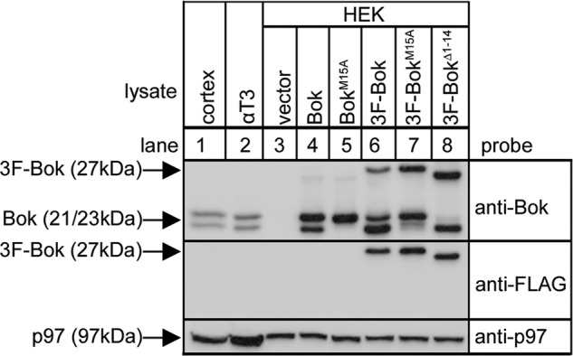
Analysis of the origin of Bok variants. Lysates from mouse brain cerebral cortex and αT3 cells (lanes 1 and 2) and HEK cells transfected to express the constructs indicated (lanes 3–8) were subjected to SDS-PAGE and probed with anti-Bok, anti-FLAG and anti-p97 as a loading control.