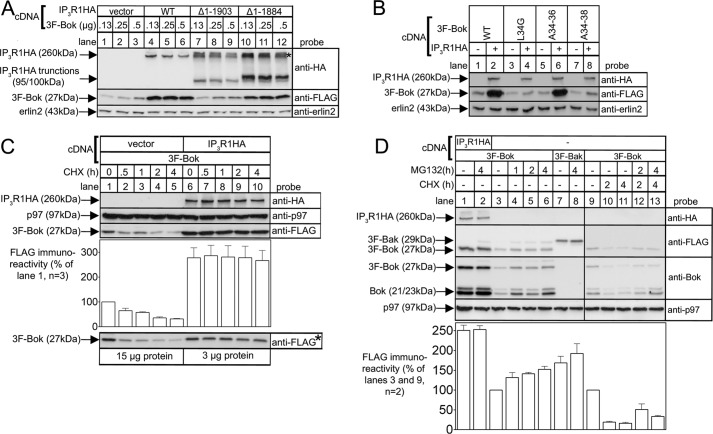
FIGURE 4.
Exogenous Bok expression is enhanced by IP3R1 and is degraded by the ubiquitin-proteasome pathway when not bound to IP3R1. HEK cells were transfected to express various 3F-Bok and/or IP3R1HA constructs, were incubated without or with 20 μg/ml CHX or 10 μm MG132 for the times indicated, were lysed, and were subjected to SDS-PAGE and probed as indicated. Erlin2 and p97 served as loading controls. The histograms show combined quantitated anti-FLAG immunoreactivity from multiple independent experiments. The asterisk in panel A marks the position in lanes 7–12 of high molecular mass (ubiquitinated) species derived from the truncated IP3R1HA constructs. The asterisked image in panel C was obtained by loading 15 μg and 3 μg of protein from vector- and IP3R1HA-transfected cells, respectively, to demonstrate that the signal from IP3R1HA-transfected cells is not saturated.

