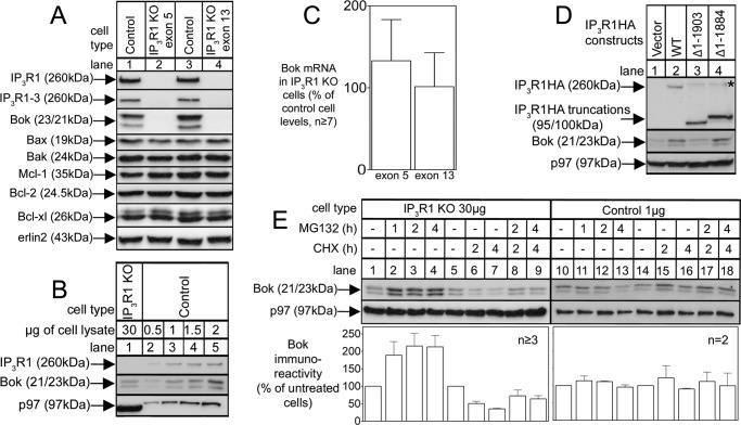
FIGURE 6.
Endogenous Bok is stabilized by IP3R1. A, levels of IP3R1 and Bok and other pertinent proteins in lysates from αT3 control and IP3R1KO cells obtained by targeting exon 5 (lane 2) and exon 13 (lane 4). B, comparison of Bok immunoreactivity in αT3 control and IP3R1KO cells obtained by loading different amounts of cell lysate. C, Bok mRNA levels (normalized to peptidylprolyl isomerase A) in exon 5 and exon 13 targeted αT3IP3R1KO cells, relative to the amount present in αT3 control cells. D, αT3 IP3R1KO cells were transfected with cDNAs encoding the IP3R1HA constructs indicated and cell lysates were probed for Bok. The asterisk marks the same species described in Fig. 4. E, αT3 control and IP3R1KO cells were treated as indicated with 10 μm MG132 and 20 μg/ml CHX, and 1 μg and 30 μg, respectively, of cell lysates were probed for Bok. The histogram shows combined quantitated Bok immunoreactivity normalized to levels in untreated cells from multiple independent experiments. An exon 5-targeted clone was used for the experiments shown in panels B, D, and E, and erlin2, and p97 served as loading controls.

