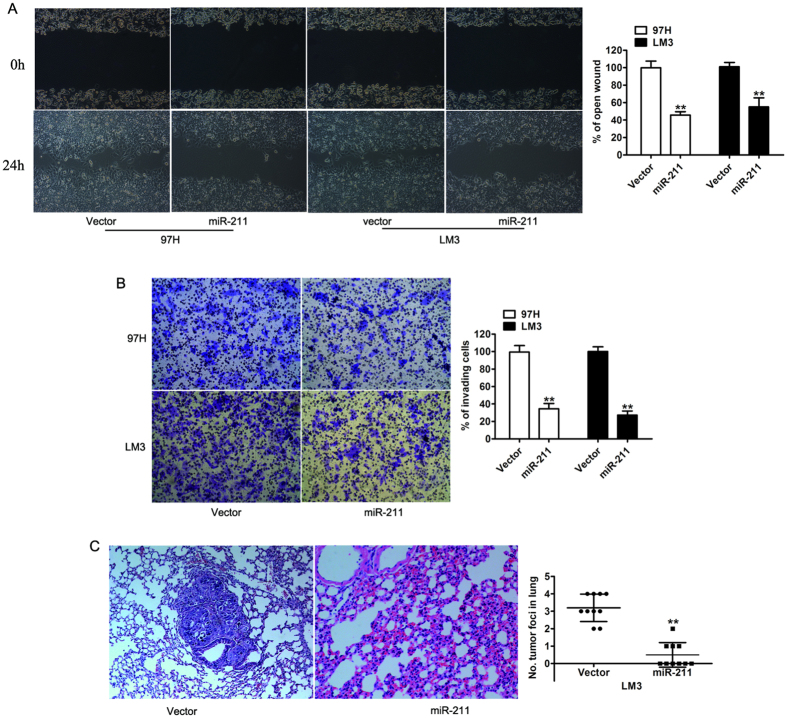
Figure 3. miR-211 suppressed tumor migration and invasion in vitro and in vivo.
(A) Representative micrographs of wound length at 0 and 24 h after wounding. The indicated cells transfected with either vector or miR-211 (left) and a histogram showed the rate of front migration of cells of 5 randomly selected fields. (B) Representative photographs of Matrigel invasion assays at 36 hours after seeding. The indicated cells transfected with either vector or miR-211 (left) and quantification of indicated invading cells in 5 random fields. (C) Representative images of lung metastases in nude mice by tail vein injection were confirmed by H&E staining. Data are presented as the mean ± SD based on three independent experiments. **P < 0.01.

