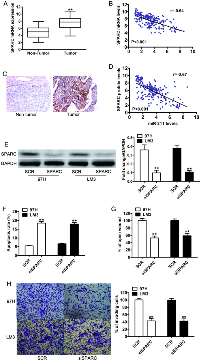
Figure 5. SPARC was significantly upregulated in HCC tissues, and its expression was inversely correlated with miR-211 expression.
(A) The mRNA expression of SPARC in 227 pairs of HCC tissues (Tumor) and their corresponding adjacent non-cancerous tissues (Non Tumor). (B) A statistically significant inverse correlation between miR-211 and SPARC mRNA levels in the 227 cases of HCC tissues. U6 and GAPDH were used as an internal control. (C) Representative images of low SPARC and high SPARC immunohistochemical staining in human HCC samples. Magnification: x100. (D) Spearman’s correlation analysis of SPARC IHC and the expression level of miR-211. Corresponding P values analyzed by t-test or Spearman correlation test were as indicated. (E) Western blot analysis of SPARC in 97H and LM3 cells transfected with scramble sequence (SCR) or siSPARC. (F) Apoptosis analysis of 97H and LM3 cells transfected with scramble sequence (SCR) or siSPARC. (G) Wound healing assay and invasion assay (H) were analyzed in 97H and LM3 cells transfected with scramble sequence (SCR) or siSPARC. **P < 0.01.

