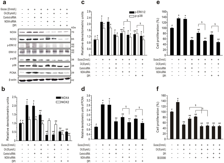
Figure 8. CA inhibits GMC proliferation mediated by NADPH/ERK1/2 and p38 MAPK signaling pathways.
(a–e) CA inhibits GMC proliferation via NADPH. GMCs serum-starved for 24 h were transiently transfected with NOX4 or control siRNA; 48 h later, the cells were pretreated with NADPH oxidase inhibitors DPI (10 μmol/L) for 30 min, followed by incubation with normal glucose (NG) or high glucose (HG) in the presence or absence of CA (30 μmol/L). To assess the inhibitory effect of CA on protein expression or cell proliferation, GMCs were further incubated for 24 h or 48 h, respectively. Representative western blots and statistical analysis are shown in (a–d). (e) Cell proliferation was measured by MTT assays. (f) CA inhibits GMC proliferation co-regulated by NADPH and p38 MAPK. GMCs were pretreated with DPI (10 μmol/L) and/or SB203580 (20 μmol/L) for 30 min, followed by treatment with NG or HG in the presence or absence of CA (30 μmol/L) for 48 h. Cell proliferation was measured by MTT assays (n = 6). *P < 0.05 vs. NG control; #P < 0.05 vs. HG control; §P < 0.05 vs. inhibitor/siRNA without CA.

