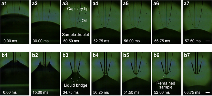
Figure 2. Series of images captured by a high-speed CCD camera in the spontaneous injection process of a capillary probe from a droplet sample.
(a) Capillary probe with hydrophobic outer surface on the tip end; (b) Capillary probe with hydrophilic outer surface on the tip end. Conditions: tapered-tip capillary, 50 μm i.d.; sample droplet, 1.0 × 10⁻² M fluorescein solution; capillary tip removing speed, 1000 mm/min.

