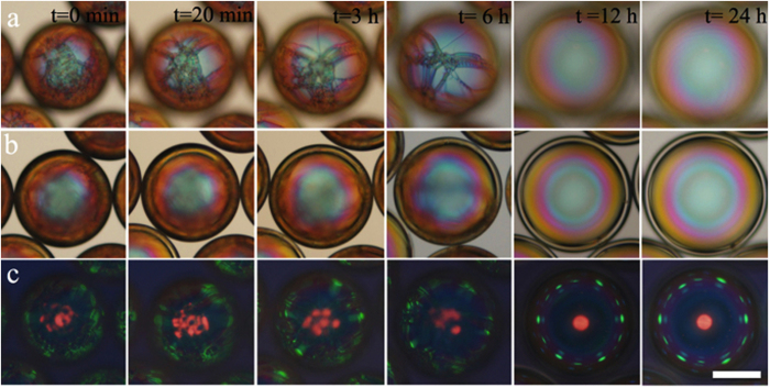
Figure 2. Removal of oily streak defects by osmotic thinning.
Optical microscopy textures in transmission without analyser (a,b; focus on top and centre plane of shell, respectively) and in reflection between crossed polarisers (c) of a cholesteric shell subjected to expansion and thinning by osmosis. The scale bar is 100 μm. After some 12 hours of osmotic expansion the shell is free of visible defects, as compared to the week-long annealing required without osmosis.

